Nusinersen

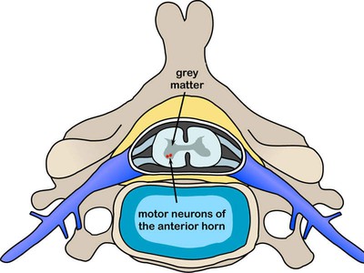
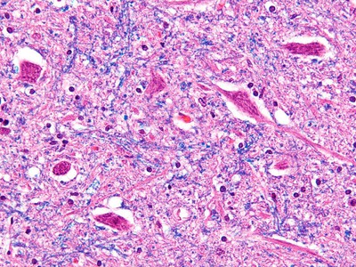
SMA(spinal muscular atrophy)는 spinal cord의 motor neuron의 loss로 인해 생기는 병이다.

fullscreen fullscreen_mobile
Spinal muscular atrophy (SMA) is caused by the loss of motor neurons in the spinal cord. Image courtesy of DO11.10.
fullscreen fullscreen_mobile
The loss of motor neurons occurs due to a defect in the SMN1 gene. Image courtesy of Nephron.
fullscreen fullscreen_mobile
Spinraza increases survival motor neuron (SMN) protein in infants and children with SMA. Image courtesy of Emw.
fullscreen fullscreen_mobile
Intron retention as a mechanism for DNA damage induction. Model for splicing dysregulation as a modulator of R-loop formation and DNA damage. Depletion of proteins in green induces DNA double-strand breaks, and depletion of proteins in blue induces R-loop formation.

Intron retention은 plant와 budding yeast에서는 gene expression regulation의 major mechanism으로 알려져 있었다. 최근까지도 mammalian system에서는 intron retention의 functional significance에 대해서는 의문점들이 있었다. 실험실에서 intron retention이 된 transcript를 찾는 등의 증거물을 확보하는 일은 기술적으로 어려운 일이었고, noise 정도의 수준으로 생각되는 정도였다. High-throughput deep sequencing 등의 기술의 발달도 정보가 넘쳐흐르자, physiological / pathological context에서 intron retention의 패턴에 대해 더 상세하게 분석할 수 있게 되었다.

정상적인 발달은 물론이고 스트레스나 질병에 대한 생체의 대응방식의 중요한 구성요소임이 최근 연구에서 밝혀졌다. 또한 intron retention이 어떠한 메커니즘으로 다음을 조절하는지 연구가 되고 있다.

  • protein isoform production
  • RNA stability and translation efficiency
  • rapid induction of expression via post-transcriptional splicing of retained introns.

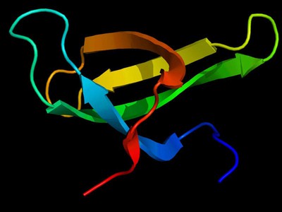
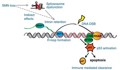
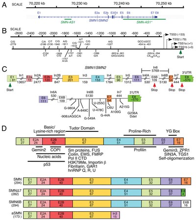
Both survival of motor neuron (*SMN*) genes are associated with spinal muscular atrophy; mutations in *SMN1* cause the disease, and *SMN2* modulates its severity

Alternative splicing of a cryptic exon embedded in intron 6 of *SMN1* and *SMN2*

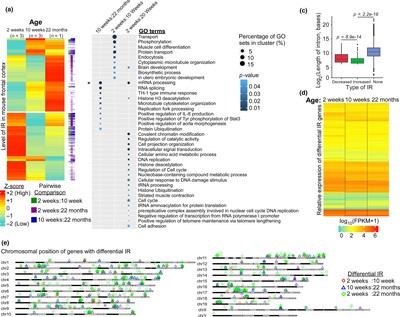
people are studying intron retention in mice brains, and the results are as follows:

fullscreen fullscreen_mobile
Intron retentions across three age‐groups in mouse frontal cortex. (a) Expression heatmap of differentially retained introns across three age‐groups (2, 10 weeks, and 22 months) in mouse frontal cortex. (b) Gene ontology analysis of the differential IR genes determined through pairwise comparison between two distinct time‐points (*p*‐value <0.05, Fisher's exact test; **p*‐value <0.05, Fisher's exact test with Benjamini–Hochberg correction). (c) Boxplot for the length distribution of the differentially retained and spliced introns. The expression level of intron at 2 weeks was used as the reference to determine the decreased or increased IR during aging. “None” refers to spliced introns (*p*‐value <0.05, Welch's *t* test). (d) Relative expression of IR genes between different ages of mouse frontal cortex as represented by Log10 (FPKM + 1) values. (e) Ideogram displaying the distribution of the differential IR genes across the mouse genome

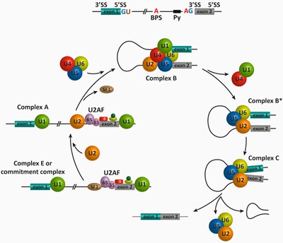
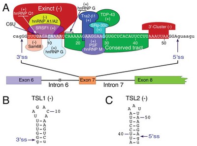
Splicing을 target하는 방법은 간단하다. 다른 diagram을 보자:

fullscreen fullscreen_mobile
Schematic representation of the spliceosome assembly and pre-mRNA splicing. In the first step of the splicing process, the 50 splice site (GU, 50 SS) is bound by the U1 snRNP, and the splicing factors SF1/BBP and U2AF cooperatively recognize the branch point sequence (BPS), the polypyrimidine (Py) tract, and the 30 splice site (AG, 30 SS) to assemble complex E. The binding of the U2 snRNP to the BPS results in the pre-spliceosomal complex A. Subsequent steps lead to the binding of the U4/U5–U6 tri-snRNP and the formation of complex B. Complex C is assembled after rearrangements that detach the U1 and U4 snRNPs to generate complex B*. Complex C is responsible for the two trans-esterification reactions at the SS. Additional rearrangements result in the excision of the intron, which is removed as a lariat RNA, and ligation of the exons. The U2, U5, and U6 snRNPs are then released from the complex and recycled for subsequent rounds of splicing.

SMN deficiency는 SMA(spinal muscular atrophy)의 severe model에서 widespread한 intron retention과 DNA damage를 일으킨다고 한다.

fullscreen fullscreen_mobile
wow
fullscreen fullscreen_mobile
mechanism2

This is really cool.

References

Mohini Jangi et al., “SMN deficiency in severe models of spinal muscular atrophy causes widespread intron retention and DNA damage”, PNAS March 21, 2017 114 (12) E2347-E2356; first published March 7, 2017 https://doi.org/10.1073/pnas.1613181114

Jacob, A.G. & Smith, C.W.J. “Intron retention as a component of regulated gene expression programs”, Hum Genet (2017) 136: 1043. https://doi.org/10.1007/s00439-017-1791-x

Satomi Yoshimoto et al., “Alternative splicing of a cryptic exon embedded in intron 6 of SMN1 and SMN2”, Hum Genet Var (2016) 3: 16040. https://doi.org/10.1038/hgv.2016.40

Swarnaseetha Adusumalli et al., “Increased intron retention is a post‐transcriptional signature associated with progressive aging and Alzheimer’s disease”, IFAA, First published: 13 March 2019 https://doi.org/10.1111/acel.12928

Marc Suñé-Pou et al., “Targeting Splicing in the Treatment of Human Disease”, Genes 2017, 8, 87; https://doi.org/10.3390/genes8030087

Wurster CD, Ludolph AC. Nusinersen for spinal muscular atrophy. Ther Adv Neurol Disord. 2018;11:1756285618754459. Published 2018 Mar 13. doi:10.1177/1756285618754459

Other Links:

https://www.drugdevelopment-technology.com/projects/spinraza-nusinersen-for-the-treatment-of-spinal-muscular-atrophy-sma/